40 years of helping those
affected by CRPS

Living With CRPS
RSDSA gives those affected by CRPS education, advocacy and hope! From signs & symptoms, to diagnosis, to living with CRPS long term, this section has health and lifestyle information for adults and youth living with this painful and debilitating condition – as well as for their families and caregivers.

Research
RSDSA raises funds for research to find better treatments – and a cure – for CRPS. We also work with research groups and healthcare professionals worldwide to foster collaboration, communication and awareness. Here you’ll find the latest studies and articles about progress and breakthroughs

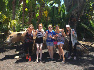
Community
CRPS can be isolating. RSDSA ensures that those affected by CRPS are not alone. We’ve built a strong, vibrant community that participates in conferences, fundraising events, mentoring, support groups and other activities that help them take control of life with CRPS. Join us.
Our Mission
Reflex Sympathetic Dystrophy Syndrome Association (RSDSA) mission is to provide support, education, and hope to all affected by the pain and disability of CRPS, while we drive research to develop better treatments and a cure.

Upcoming Events
Abbott is hosting free national patient education webinar events in 2024.
RSDSA's 2024 Young Adult Retreat will take place in Denver, Colorado from Friday, May 31st to Monday, June 3rd!
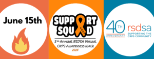
This is our 5th Virtual Walk as well as a celebration of RSDSA's 40th Anniversary!
Corporate Partners


Recent Videos
5th Annual RSDSA Long Island CRPS Awareness Walk & Expo Radio Interview